CT Scan vs. MRI: What’s the Difference?
CT (computed tomography) and MRI (magnetic resonance imaging) are both used to diagnose and stage cancer. Many people do not know the difference between the two methods or why one might be selected over the other.
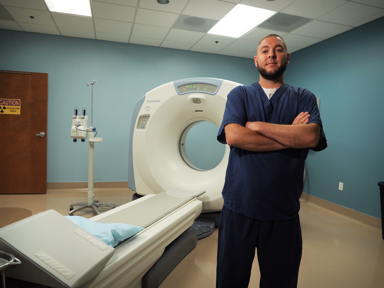
What does a CT scan show?
A CT scan uses X-rays to create detailed pictures of organs, bones, and other tissues. The person lies on a table that moves through a scanning ring, which looks like a large doughnut. The data collected can be assembled to form three-dimensional images. The images reveal abnormalities in both bone and soft tissues, such as pneumonia in the lungs, tumors in different organs, or bone fractures.
Some of the common things a CT scan is used for are:
- Bone fractures
- Tumors
- Cancer Monitoring
- Internal Bleeding
What does an MRI show?
MRI also creates detailed pictures of areas inside the body, but it uses radio waves and a powerful magnet to generate the pictures. The person also lies on a table that moves into a doughnut-shaped device, but the doughnut is much thicker. Similarly, these pictures can show the difference between normal and diseased tissue.
Here are some common areas of the body an MRI diagnose:
- Joint
- Brain
- Wrist
- Ankle
- Breast
- Heart
- Blood Vessels
What are the advantages of a CT scan?
With a CT scan, we can create an image of almost the entire body, from the neck to the thighs, in a few seconds. CTs are incredibly useful for diagnosing and staging cancer, checking whether it has come back, and monitoring whether a treatment is working. It’s very effective for surveying the entire body to look for places where the cancer has spread, such as the lungs, liver, or bone. These are called metastases. Most of the time, CT is the first choice to stage cancer. Apart from cancer, CT scans are often used to image bone fractures and look for internal bleeding or blood clots, spinal and brain injuries, and other conditions.
What are the advantages of MRI?
Where MRI really excels is showing certain diseases that a CT scan cannot detect. Some cancers, such as prostate cancer, uterine cancer, and certain liver cancers, are pretty much invisible or very hard to detect on a CT scan. Metastases to the bone and brain also show up better on an MRI. This imaging is also used for many purposes unrelated to cancer, including injuries to soft tissue or joints, and injury or disease of internal organs including the brain, heart, and digestive organs.
What are some disadvantages of each imaging method?
Because CTs use ionizing radiation, they could damage DNA and may very slightly raise the risk of developing cancer. The U.S. Food and Drug Administration estimates that the extra risk of any one person developing a fatal cancer from a typical CT procedure is about 1 in 2,000. MRIs do not use ionizing radiation, so there is no issue of raising cancer risk. But they take much longer to complete than CTs. MRIs require the person to lie still within a closed space for about 20 to 40 minutes. This can affect some people with claustrophobia, and the procedure is noisy, which is why we provide ear protection.
Both CT and MRI commonly require the injection of a contrast dye before or during the procedure. This helps the radiologist see organs and other tissues within the body more clearly.
What concerns do people have about either imaging method?
For CT, We hear anxiety about exposure to radiation, especially if it is being done repeatedly. For example, certain early-stage cancers can be cured. But you might be coming back every few months or every year for a CT scan. The question is: Do we have an alternative? For detecting cancer that has come back throughout the body, a CT scan is preferable to an MRI. As radiologists, we follow a measure called “as low as reasonably achievable.” This means we give enough radiation to create CT images that are of high enough quality that we can make a good clinical decision, but we keep the radiation as low as possible to minimize risk.
For MRI, people who have trouble with claustrophobia or are unable to hold their breath, which may be required for certain abdominal imaging tests, may not be able to tolerate the procedure. Some MRI machines can be configured in ways that may reduce claustrophobia. Medical implants, such as a pacemaker, brain stimulator, or other devices, are another complicating factor. The radio waves used with MRI can heat up devices made of metal. This is potentially a concern for something inside the body. Newer medical devices are usually designed with this in mind, so they are safe inside an MRI.
How do doctors decide which imaging a person should receive?
We usually use CT first for most people, unless a tumor is much better seen on MRI. But we go back and forth as needed. If we see something on a CT scan we’re unsure about, we may recommend an MRI for further evaluation. If someone has several MRIs and is unable to lie still or hold their breath so we can get a good image, we may suggest a CT as an alternative. We’re guided by the principle of whether the benefits of a test outweigh its risks. That’s what medical imaging is about.
CT scan (computed tomography) and MRI (magnetic resonance imaging) are two types of medical imaging tests used to help diagnose and evaluate different medical conditions. While both tests provide doctors with detailed images of the body's internal structures, there are several key differences between CT scans and MRIs, including:
- Technology: CT scans use X-rays and a computer to create detailed cross-sectional images of the body. MRIs use a strong magnetic field and radio waves to create detailed images of the body's internal structures.
- Radiation exposure: CT scans expose patients to a small amount of ionizing radiation, which can increase the risk of cancer with frequent exposure. MRIs do not use ionizing radiation and are considered safer in this regard.
- Image quality: CT scans are better suited for imaging bone and lung tissue because they provide detailed images of these dense structures. MRIs are better suited for soft tissue imaging, such as the brain, spinal cord, and joints.
- Time: CT scans are typically faster than MRIs, taking only a few minutes to complete. MRIs can take up to an hour or more to complete.
- Cost: CT scans are generally less expensive than MRIs.
Ultimately, the choice of imaging test depends on the patient's condition, the part of the body being imaged, and the doctor's preference. In some cases, both CT scans and MRIs may be used together to provide a more comprehensive diagnosis.
Access 24/7 Imaging Service
A physician can help determine the correct procedure for you. At ER of Texas emergency room, our state-of-the-art onsite laboratory facility, matched with our expert staff, provides advanced imaging services. Discover 24/7 access to CT Scans that get you the results you need faster, more accurately, and efficiently.
We have 9 facilities spread across the DFW area that are OPEN 24/7 located in Hurst, Colleyville, Frisco, Highland Village, Hillcrest, Uptown, Little Elm, Mansfield, and Texoma.
.jpg)